A heart valves contouring atlas on average intensity projection 4D-CT for lung cancer radiotherapy
PO-1177
Abstract
A heart valves contouring atlas on average intensity projection 4D-CT for lung cancer radiotherapy
Authors: JOANNA SOCHA1, Anna Rygielska2, Beata Uziębło-Życzkowska3, Justyna Chałubińska-Fendler1, Agnieszka Jurek3, Małgorzata Maciorowska3, Marta Mielniczuk3, Paweł Pawłowski1, Dobromira Tyc-Szczepaniak1, Lucyna Kępka1
1Military Institute of Medicine, Department of Radiotherapy, Warsaw, Poland; 2Military Institute of Medicine, Department of Radiotherapy, Laboratory of Medical Physics, Warsaw, Poland; 3Military Institute of Medicine, Department of Cardiology and Internal Diseases, Warsaw, Poland
Show Affiliations
Hide Affiliations
Purpose or Objective
A
detailed contouring atlas of the heart valves is lacking. Existing heart
contouring atlases have not been evaluated on average intensity projection four-dimensional
noncontrast computed tomography (AVE 4D-CT) scans, routinely used for
organ-at-risk delineation in lung cancer radiotherapy (RT). As a first step of
a planned prospective trial on imaging-based evaluation of RT-related
cardiotoxicity in NSCLC, we aimed to develop the heart valves contouring atlas
and to assess interobserver variation in delineation of the heart, its substructures,
and coronary arteries on AVE 4D-CT scans, along with the impact of contour
variation on estimated RT doses.
Material and Methods
A draft
of the heart valves contouring atlas with written guidelines was developed by a
radiation oncologist (JS) and agreed by cardiologists. Five radiation
oncologists and four cardiologists were recruited to delineate the valves
according to the draft, and the remaining heart substructures (4 chambers, 4 left ventricle segments, 4 coronary arteries and the
heart) based on the existing
heart contouring atlases on AVE 4D-CT scans of ten patients who underwent
radio(chemo)therapy for NSCLC. Based on the delineation exercise, some modifications of the initial
draft of the heart valves atlas were made and their delineation was repeated. Next,
the "reference" contours for each structure and case were
collectively defined, to which all the observer contours were compared. Spatial variation was
assessed using the Sørensen-Dice similarity coefficient (DSC) and the directed
average Hausdorff distance (DAH). The effect of spatial
variation on RT doses was assessed using the patients' RT treatment plans. The
results presented below are for the patient with the worst reproducibility.
Results
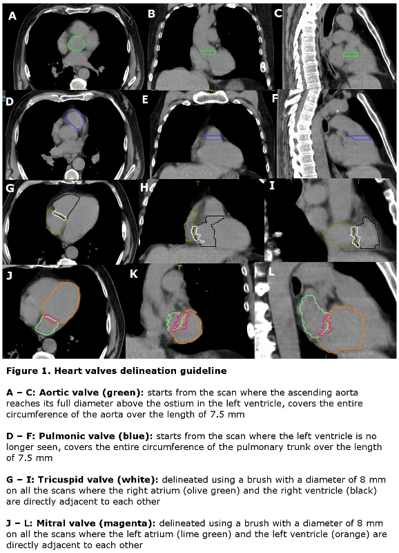
A detailed contouring atlas of the heart valves was
developed (Fig.1) and evaluated on AVE 4D-CT scans: inter-observer
contour overlap (mean DSC) was 0.62, 0.55, 0.43 and 0.36, and interobserver
contour separation (mean DAH) was 2.3, 3.4, 2.7 and 3.6 mm for the pulmonic, aortic,
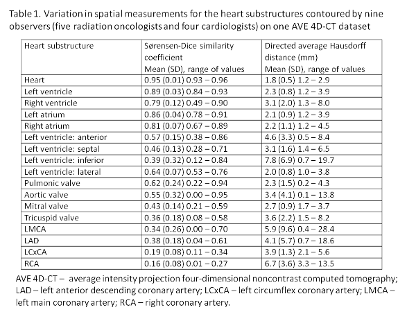
mitral and tricuspid valve, respectively. For the heart and its remaining
substructures, defined according to the existing atlases, the interobserver agreement
was the highest for the heart and its four chambers, lower for the left
ventricular segments and the lowest for the coronary arteries (Tab.1). In the
presented patient, this spatial variation resulted in <1.5Gy dose variation for
11 of 17 heart substructures contoured by nine observers on one AVE 4D-CT
dataset; the highest variation resulting in the difference of 7.64Gy between
the minimum and maximum estimated dose was recorded for the left main coronary
artery.
Conclusion
Our atlas
enables reproducible delineation of the heart valves. Delineation of the heart and
its substructures on AVE 4D-CT scans according to the existing atlases is
feasible, with inter-observer variability similar to that reported in
validation studies of these atlases on conventional noncontrast CT scans.