Validation of optical surface/thermal imaging and X-ray positioning accuracy for SRS treatments
Vanessa da Silva Mendes,
Germany
PD-0781
Abstract
Validation of optical surface/thermal imaging and X-ray positioning accuracy for SRS treatments
Authors: Vanessa da Silva Mendes1, Michael Reiner1, Stefanie Corradini1, Maximilian Niyazi1, Claus Belka1, Guillaume Landry1, Philipp Freislederer1
1University Hospital, LMU Munich, Department of Radiation Oncology, Munich, Germany
Show Affiliations
Hide Affiliations
Purpose or Objective
The novel Exactrac Dynamic (Brainlab AG,
Germany) provides real-time 3D surface imaging by combining structured light
with thermal information, and is complemented by an in-room kV X-ray imaging
system. The aim of the study was to compare the
positioning accuracy of the combined surface/thermal imaging system and the
gold-standard, stereoscopic X-rays, for stereotactic radiosurgery (SRS)
treatments. In order to provide a more realistic scenario, a head phantom
prototype with a specific heat signature profile was used. Phantom
specifications regarding surface temperature stability were also investigated.
Material and Methods
An anthropomorphic 3D-printed
head phantom (Prime, RTsafe, Greece) with bone equivalent material, 3 embedded
ball bearings and can be filled with water up to 45°C was used. It was fixed to the
table using a 4Pi open face mask (Brainlab AG, Germany). To investigate the phantom’s surface
temperature stability, the phantom was filled with warm water (41°C) and surface
temperature was measured with an infrared thermometer at 7 locations within the
area of the face opening, app. every 10 min, for 65 min. The surface/thermal imaging positioning (DS+T)
was measured and compared to X-ray based positioning (DX-RAY) at an
Elekta Versa HD linac. The couch (at 0°) was displaced in all 3 directions (lateral,
longitudinal and vertical) from the planned position (10 random translations,
max. translation 2.5 mm), using the HexaPODTM evo RT interface
(Elekta AB, Sweden). An empty (cold) phantom at room temperature and a warm
water filled phantom were used. The difference between both positioning methods
(ΔDPOSITION=ǀDX-RAY-DS+Tǀ) was
calculated and an independent t-test was done to investigate the significance
of the differences for a cold and a warm phantom.
Results
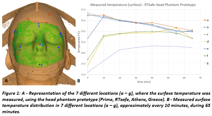
Fig. 1 shows the temperature curves, where a stabilisation
is seen 10 min after filling the phantom and during the next 55 min. In 6 out
of 7 locations values measured within nearly 1h were between 36°C and 32°C. The
4°C difference is not expected to impact the measurements as the surface/thermal
imaging reference is updated and zeroed after X-rays are acquired.
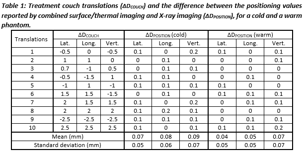
Tab. 1 shows the couch translations and the difference between both positioning methods. No statistical significance between the positioning of the cold and warm phantom was found (p
˃ 0.05).
Conclusion
Considering the surface temperature stability this
phantom demonstrated to be suitable for measurements with ExacTrac Dynamic for
at least 1h. The measurements with a warm phantom showed good
agreement between surface/temperature and
X-ray imaging. No deviations larger than 0.1 mm were found, leaving open the
possibility of monitoring the patient using more surface guidance and less
ionising radiation for cranial treatments. Measurements with a
cold phantom showed slightly higher deviations. However, as the differences
were not statistically significant, a larger set of measurements is planned to
determine whether a heated phantom is essential for SRS QA.