Intra-mandible radio-sensitivity for osteoradionecrosis: effect of local dose and teeth extractions
Nienke Sijtsema,
The Netherlands
PO-1820
Abstract
Intra-mandible radio-sensitivity for osteoradionecrosis: effect of local dose and teeth extractions
Authors: Nienke Sijtsema1,2, Gerda Verduijn1, Yvette van Norden1, Hetty Mast3, Aad van der Lugt2, Mischa Hoogeman1,4, Steven Petit1
1Erasmus MC Cancer Institute, Department of Radiotherapy, Rotterdam, The Netherlands; 2Erasmus MC, Department of Radiology and Nuclear Medicine, Rotterdam, The Netherlands; 3Erasmus MC, Department of Oral and Maxillofacial Surgery, Rotterdam, The Netherlands; 4HollandPTC, Department of Medical Physics and Informatics, Delft, The Netherlands
Show Affiliations
Hide Affiliations
Purpose or Objective
Osteoradionecrosis (ORN) of the mandible is a severe late effect of radiotherapy
for head and neck tumors. It is most often observed in premolar and molar
regions, which suggest not all mandibular regions are equally prone for
development of ORN. Therefore, we investigated the interaction between the ORN
location, the dose to that location and the location of teeth extractions in a
group of 324 oropharyngeal squamous cell carcinoma patients treated with
(chemo)radiotherapy.
Material and Methods
For patients with ORN after radiotherapy, the ORN volume was delineated
on the planning CT. All dose distributions were converted to 2 Gy equivalent
dose based on α/β=3 Gy, accumulated, and deformed non-rigidly to
a reference patient using ADMIRE 3.7.7 (Elekta AB, Stockholm, Sweden). The
reference patient contained delineations of 16 mandible subregions
corresponding to the 16 dental elements. Multi-variable logistic regressions were
performed per mandible subregion to relate the presence of ORN in a subregion
to its mean dose, whether the dental element in the subregion was extracted and
whether its neighbor elements (i.e. other dental elements on the same side of the mandible) were extracted. Likelihood ratio tests were
performed to compare models, with p≤0.05
considered statistically significant. In the first regression all dental elements
from all patients were combined. Next, separate regression analyses were
performed per subregion (left and right combined).
Results
Patients could be registered accurately to the reference patient. The 95
percentile of the distance between the mandible delineations was on average 3.50
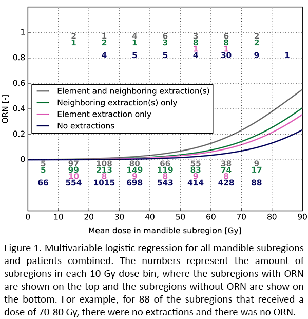
mm (range 1.51-8.12 mm). 27 Patients developed ORN (8%). In the combined analysis
the mean dose to the mandible subregion, pre-radiotherapy teeth extraction in the mandible subregion, and
pre-radiotherapy teeth extractions of neighboring dental elements were all
associated with an increased risk of ORN (Figure 1). The odds ratios were 1.073 per unit increase in Gy (95% confidence
interval (CI): (1.059,1.088)), 1.79 (95%
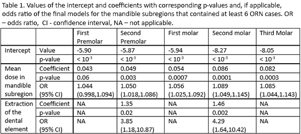
CI: (1.023,3.11)), and 2.24 (95% CI: (1.39,3.54)), respectively. In the analysis per mandible subregion (Table
1) the dose was associated with an increased risk of ORN in the second
premolar, first molar, second molar, and third molar. Extractions were
associated with an increased risk of ORN only in the second premolar (p-value =
0.03) and in the second molar (p-value = 0.004). Due to limited statistical
power, extraction of neighboring dental elements was not taken into account in
the regressions per subregion.
Conclusion
Large intra-mandible differences in radio-sensitivity for ORN were
observed. Dose effect relations were established for different regions of the
mandible corresponding to dental elements. Extraction of the dental element of
interest or its neighbors increased the local sensitivity of the mandible for
ORN. These findings strongly support, and can guide studies to selectively
spare sensitive regions of the mandible to decrease the risk of ORN.