Quality assurance of electronic brachytherapy treatment units with a plastic scintillation detector
PH-0325
Abstract
Quality assurance of electronic brachytherapy treatment units with a plastic scintillation detector
Authors: Peter Georgi1, Gustavo Kertzscher1, Thorsten Schneider2, Jacob Graversen Johansen1, Kari Tanderup1, Lars Nyvang1
1Aarhus University Hospital, Department of Oncology, Aarhus, Denmark; 2Physikalisch-Technische Bundesanstalt, Radiation Protection Dosimetry, Braunschweig, Germany
Show Affiliations
Hide Affiliations
Purpose or Objective
Standardized quality
assurance (QA) methods are limited in electronic brachytherapy (eBT) [1]. The
purpose of this experiments was to design a simple and accurate method for verification of the relative absorbed dose to water distribution. For the first
time, the dose distribution from an eBT
source was measured in water using a small plastic scintillation detector
(PSD).
Material and Methods
The Papillon 50
(P50; Ariane Medical Systems Ltd, UK) is an eBT source mainly used for rectal
cancer treatment. It delivers 50 kVp X-rays (half value layer ~ 0.7mmAl). The
beam is collimated using cylindrical steel applicators (Ø22-30 mm). The absorbed dose from the P50 source, with a Ø25
mm applicator, was measured with a PSD. The system consisted of a cylindrical
plastic scintillator (Ø1 mm, L=0.5 mm) coupled to an optical fibre which
transmitted the scintillation light to a photo multiplier tube (PMT) (H5783
SEL3, Hamamatsu). The PMT was coupled to an electrometer (Unidos Webline, PTW
Freiburg Germany). The PSD was placed on a motorised stage in a water phantom (MP3,
PTW), while the P50 applicator was pointed vertically downward, with the tip
just breaching the water surface of the phantom. The applicator was rigidly
fixed in a custom build frame. The PSD was moved to predetermined positions where
the relative dose rate was measured. The movement and measurements were
automated with the software Mephysto (PTW). The dose depth curves and dose
profiles at various depths were measured in steps of 0.5-5 mm. The width of the
dose profiles at 50% of the central dose was determined.
Results
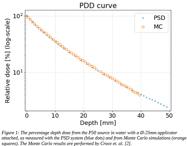
Depth-dose curves:
The measured percentage depth dose (PDD) shows an
almost exponential decay with 1-order reduction every 25 mm (fig. 1). The
measurements are in good agreement with Monte Carlo (MC) simulations performed
by Croce et. al. [2].
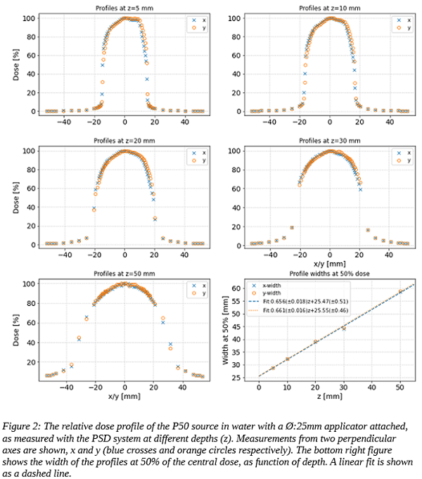
Dose profiles:
The dose profile smears out and the width increases for
larger depths (fig. 2). The increase follows a linear behavior (dashed lines
in fig. 2 bottom right). There is no significant directional dependency. The
z=5, 10, and 20 mm profiles are asymmetric, with a shoulder to the right,
likely due to the X-ray rod not being exactly centered inside the applicator.
Conclusion
The PDD and dose
profiles, at various depths, of an eBT source have been measured under full
scatter conditions in a water phantom for the first time with high spatial
resolution. The measurements were performed with a novel PSD system, which
offers a simple method for QA of the relative absorbed dose to water of eBT treatment units. The method can
easily be transferred to other low energy X-ray sources.
References:
[1] Primary Standards and Traceable Measurement Methods for X-ray
Emitting Electronic Brachytherapy Devices, Publishable Summary (2018):
[2] O. Croce, S. et al. Radiation Physics and Chemistry 81 (2012) 609-617