ESTRO 2023 Physics Track Report - ‘Big data, big headache’ joint ESTRO-American Association of Physicists in Medicine session
Dealing with follow-up images
My data was causing such a headache that venting to any colleague unlucky enough to cross my path didn’t cut it anymore, so when ESTRO contacted me about the ‘Big data, Big headache’ session at ESTRO 2023, I jumped at the chance to vent to a wider audience (Fig. 1). Despite the headache, I think we all appreciate the vital role that big data plays in improving patient outcomes. But, group therapy is always useful, so I chose to talk about my headache of working with follow-up imaging.
Follow-up imaging in radiation oncology is typically used to assess treatment response. A common requirement for follow-up imaging research is high-quality, standardised images that are captured at a specified frequency. However, this is often not the case when working with retrospective and routine data. I experienced this headache while working on a project to assess facial asymmetry through the use of routine MRI of children who have received radiotherapy for head-and-neck tumours [1].
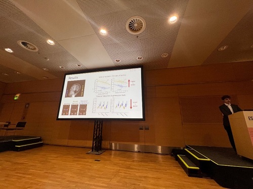
In routine follow-up of childhood cancer survivors, typically two 2D MRIs (axial and coronal) are acquired to minimise time spent on scan acquisition, and acquisition of volumetric MRI is rare. The 2D acquisition has high in-plane resolution, but low out-of-plane resolution and limited field-of-view. Such images can be used to assess treatment response, but not to perform accurate facial-asymmetry measurements. Our solution was to develop a super-resolution method to generate the high-resolution data required (Fig. 2). This enabled us to make use of routine clinical data, which is preferable to running a prospective study, as long-term follow-up (>5 years) is required in paediatric research. The clinical team provided insight into this data, which was integral to each stage of the project. The progress we made together lessened my headache, and our solutions can be summarised as follows:
- Keep up-to-date with novel approaches in image processing and artificial intelligence that might aid your research.
- Get to know your data and data acquisition pipeline.
- Work within a multidisciplinary team and get your clinicians involved!
In summary, many research projects are ambitious and the data collection can sometimes cause a big headache. But, with some novel approaches and a great multidisciplinary team, we can ‘make the most of what we have’ and still perform big-data research. I hope to see lots more projects making use of routine follow-up imaging in future!
References
[1] https://www.friendsofrosie.co.uk/reducing-the-risk-of-facial-asymmetry-after-radiotherapy/
.jpg.aspx)
Fig. 1 Presentation of ‘dealing with follow-up images’ to a full audience in the ‘Big data, Big headache’ session of ESTRO 2023.

Fig. 2 Jose Benitez Aurioles (PhD student from the University of Manchester, UK) presenting on super-resolution of paediatric MRI in the mini oral sessions.

Angela Davey
Postdoctoral research associate
Division of Cancer Sciences, University of Manchester
UK
@AngieDavey_
Angela.davey@manchester.ac.uk
About the author:
Dr Angela Davey is a postdoctoral research associate in the Division of Cancer Sciences at the University of Manchester. Her current research involves analysis of routine data to investigate ways to improve radiotherapy treatments for children with cancer and hence to reduce their risk of side effects later in life.