One of the biggest hurdles for those who treat cancer is identifying patients who are more likely to respond favourably to a given anticancer therapy. Refinement of patient selection to optimise radiation therapy benefit is highly anticipated from biological markers, including tissue-derived invasive analytes and non-invasive biological assessments, such as functional imaging or liquid biopsy biomarkers. In contrast with traditional tissue biopsies that are used in clinical practice for pathological diagnoses and to provide a static snapshot of cancer, analysis of liquid biopsies is a non-invasive approach that could be used to detect cancer early, to identify patients with metastatic disease, to monitor treatment efficiency in real-time, and to analyse the dynamic nature of cancer during the treatment, as well as to identify therapeutic targets and resistance mechanisms. However, despite extensive efforts to identify and validate various liquid biopsy analytes over the last decade, only analyses based on blood-based circulating cell-free DNA (cfDNA) and circulating tumour cells (CTCs) have been approved for routine clinical practice.1,2
cfDNA that is released into circulation is derived from non-cancerous tissues, mainly leukocytes and stromal cells, as well as from the tumour (which is termed circulating tumour DNA, ctDNA). Short-lived ctDNA provides a faithful real-time tumour snapshot because it retains genetic and epigenetic features of the original tumour, such as mutations and methylation patterns. Radiogenomics using ctDNA is a fast-emerging field within radiation oncology.3,4 There is accumulating evidence that pre-treatment ctDNA analyses and the longitudinal dynamics of cfDNA during chemoradiotherapy are predictors of disease control in patients with head and neck squamous cell carcinoma (HNSCC), including oropharyngeal cancer that is positive for human papilloma virus5,6 and nasopharyngeal cancer associated with Epstein-Barr virus,7,8 as well as oesophageal cancer,9 rectal cancer10,11 and non-small-cell lung cancer (NSCLC).12 Furthermore, we are able to identify the cfDNA cell of origin by genomic and epigenetic analyses.13 Recent studies show differential dynamics of cfDNA from tumour and normal tissues during radiotherapy of patients with breast and oesophageal cancer.14,15 These findings provide the basis for the radiotherapy intensification or diminution to make it more personalised3,15,16 The use of recently described predictive models that combine ctDNA and radiomics image analyses with tumour features in patients with NSCLC17 and locally advanced rectal cancer18,19 who are undergoing chemoradiotherapy have led to improved outcome predictions. These results prove the complementarity and clinical utility of integrating radiomics and liquid biopsy markers into real-time response-adaptive therapy.
The enumeration and molecular analysis of CTCs that are shed into circulation from primary tumours or metastases may also be used as non-invasive markers for precision management of radiation therapy.19-22 Serial analyses of CTCs during treatment provide valuable information on tumour heterogeneity at the DNA, RNA, and protein levels, although CTC isolation requires the use of complex techniques and equipment.2,23 Multiparameter analyses of CTCs for expression of therapeutic targets, such as programmed death-ligand 1 (PD-L1) and phosphorylated epidermal growth factor receptor (EGFR), as well as the radiation-induced DNA damage marker γH2AX, were also shown to be possible in patients with HNSCC, breast and prostate cancer.24,25
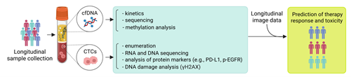
Despite this increasing evidence of the prognostic value of ctDNA and CTC analysis, the use of longitudinal liquid biopsy analyses to guide doses and schedules of radiotherapy is still at an early stage. The proof-of-concept studies have to be independently validated across cohorts, and the technical aspects of the analyses must be further developed and standardised to facilitate interpretation and clinical translation. Taken together, however, these novel technological developments provide unprecedented possibilities to employ liquid biopsy multiomics in combination with functional imaging in order to monitor radiation-induced effects in tumour and normal tissues during the treatment, and to enable the scheduling of personalised adaptive radiotherapy (Figure 1).
Figure 1. Potential application of liquid biopsy multiomics with functional imaging in order to predict therapy response and normal tissue toxicity, and to schedule adaptive radiotherapy. Created in BioRender.
Dr Ielizaveta Gorodetska (left)
OncoRay – National Centre for Radiation Research in Oncology
Faculty of Medicine, University Hospital Carl Gustav Carus, Technische Universität Dresden
Helmholtz-Zentrum Dresden – Rossendorf, Institute of Radiooncology
Germany
Professor Anna Dubrovska (right)
OncoRay – National Centre for Radiation Research in Oncology
Faculty of Medicine, University Hospital Carl Gustav Carus, Technische Universität Dresden
Helmholtz-Zentrum Dresden – Rossendorf, Institute of Radiooncology
Germany
References:
1 Saldanha et al. J Liq Biopsy 5, 100156, doi:10.1016/j.jlb.2024.100156 (2024).
2 Alix-Panabieres et al. Cancer Cell 43, 161-165, doi:10.1016/j.ccell.2024.11.009 (2025).
3 De Michino et al. Int J Radiat Oncol Biol Phys 107, 873-886, doi:10.1016/j.ijrobp.2020.05.008 (2020).
4 Earland et al. Semin Radiat Oncol 33, 262-278, doi:10.1016/j.semradonc.2023.03.004 (2023).
5 Chera et al. Clin Cancer Res 25, 4682-4690, doi:10.1158/1078-0432.CCR-19-0211 (2019).
6 Hilke et al. Radiother Oncol 151, 182-189, doi:10.1016/j.radonc.2020.07.027 (2020).
7 Lv et al. Nat Commun 10, 3941, doi:10.1038/s41467-019-11853-y (2019).
8 Lv et al. Cancer Cell 42, 1401-1414 e1404, doi:10.1016/j.ccell.2024.07.001 (2024).
9 Azad et al. Gastroenterology 158, 494-505 e496, doi:10.1053/j.gastro.2019.10.039 (2020).
10 Zhou et al. EBioMedicine 112, 105548, doi:10.1016/j.ebiom.2024.105548 (2025).
11 Clasen et al. Radiat Oncol 19, 153, doi:10.1186/s13014-024-02540-4 (2024).
12 Pan et al. Cancer Cell 41, 1763-1773 e1764, doi:10.1016/j.ccell.2023.09.007 (2023).
13 Barefoot et al. Front Genet 12, 671057, doi:10.3389/fgene.2021.671057 (2021).
14 McNamara et al. JCI Insight 8, doi:10.1172/jci.insight.156529 (2023).
15 Roth et al. Gastrointest Disord (Basel) 3, 100-112, doi:10.3390/gidisord3030011 (2021).
16 McLaren et al. Br J Cancer 129, 900-903, doi:10.1038/s41416-023-02398-5 (2023).
17 Moding et al. Cancer Discov 15, 1609-1629, doi:10.1158/2159-8290.CD-24-1704 (2025).
18 Wang et al. PLoS Med 18, e1003741, doi:10.1371/journal.pmed.1003741 (2021).
19 Wurschi et al. Medicine (Baltimore) 102, e33575, doi:10.1097/MD.0000000000033575 (2023).
20 Zapatero et al. Radiat Oncol 15, 137, doi:10.1186/s13014-020-01577-5 (2020).
21 Tinhofer et al. Ann Oncol 25, 2042-2047, doi:10.1093/annonc/mdu271 (2014).
22 Klusa et al. Int J Cancer 152, 2639-2654, doi:10.1002/ijc.34457 (2023).
23 Dai et al. Cancer Cell 43, 1399-1422, doi:10.1016/j.ccell.2025.07.008 (2025).
24 Staudte et al. Cancers (Basel) 14, doi:10.3390/cancers14112810 (2022).
25 Chatzkel et al. Future Sci OA 6, FSO437, doi:10.2144/fsoa-2019-0092 (2019).