Abstract

Title

Radiosensitizing effect of Trabectidin on human soft tissue sarcoma cells

Authors

Michele Aquilano1, Giulia Salvatore1, Mauro Loi2, Daniela Greto2, Erika Scoccimarro1, Lucia Pia Ciccone1, Giulia Stocchi1, Viola Salvestrini1, Costanza Santini1, Mariangela Sottili1, Monica Mangoni1, Lorenzo Livi3

Authors Affiliations

1University of Florence, Department of Biomedical, Experimental, and Clinical Sciences "Mario Serio", Florence, Italy; 2Azienda Ospedaliero-Universitaria Careggi, Department of Radiation Oncology, Florence, Italy; 3University of Florence,, Department of Biomedical, Experimental, and Clinical Sciences "Mario Serio", Florence, Italy

Purpose or Objective

Soft tissue sarcomas (STS) are aggressive tumors with limited effective therapeutic options. Trabectedin is indicated for the treatment of adult patients with advanced STS. The aim of the study is to evaluate in vitro if trabectedin could enhance radiotherapy by increasing cell radiosensitivity and biological aggressiveness on human cell lines of fibrosarcoma (FS), leiomyosarcoma (LMS), liposarcoma (LPS) and rhabdomyosarcoma (RS)

Materials and Methods

For each human FS (HS 93.T), LMS (HS5.T), LPS (SW872), and RS cell line (RD), IC50 was determined by colorimetric assay (MTS). Surviving Fraction (SF) was assessed at Clonogenic assay (CGA) on cells following irradiation (IR) to a dose of 2, 4 or 6 Gy, with or without trabectedin 24 h before IR to calculate radiosensitization enhancement ratio at 50 % survival (ER50). Matrigel invasion assay was performed in 4 groups:  IR 4 Gy; IR 4 Gy + trabectedin; trabectedin; control. Repartition to radiosensitive G2/ M phase, cell cycle analyses was assessed  with flow cytometry. Induction of DNA strand break and DNA repair have been observed by detecting H2AX serine 139 phosphorylation and quantifying γ-H2AX foci at 30 minutes, 6 h and  24 h after IR. 

Results

IC50 was 0,9654 nM; 0,6836 nM;1,296 nM;0,8549 nM for RS,LPS,LMS and FS respectively. Significant reduction of SF in LMS and LPS cell lines was observed following IR+trabectedin as compared to IR alone, resulting in ER50 of 2.35 and 1.45, respectively. (Fig.A)
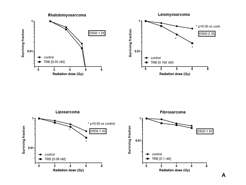
All STS cell lines showed a significantly reduced invasiness following trabectedin alone or trabectedin+IR compared to control and trabectedin+IR compared to IR alone. Trabectedin+IR compared to control, resulted in an increasing, similar or reduced G2/M phase cell fraction of cells in LPS, FS and RS/LMS, respectively. In all STS cell lines, trabectedine+IR induced a significantly higher occurrence of γ-H2AX foci compared to control, trabectedin and IR alone. Reduction in the fluorescence intensity associated to the number of foci over 24 hour was significantly lower in the combined treatment arm.


Conclusion

IC50 of Trabectedin for different STS cell line was calculated. Trabectedin+IR induced significant SF reduction in LMS and LPS cell lines and decreased cell invasiveness in all cell lines compared to IR alone. Variable effects on cell cycle were observed according to STS subtype. Trabectedin + IR resulted in increased γ-H2AX foci formation and impaired damage repair.